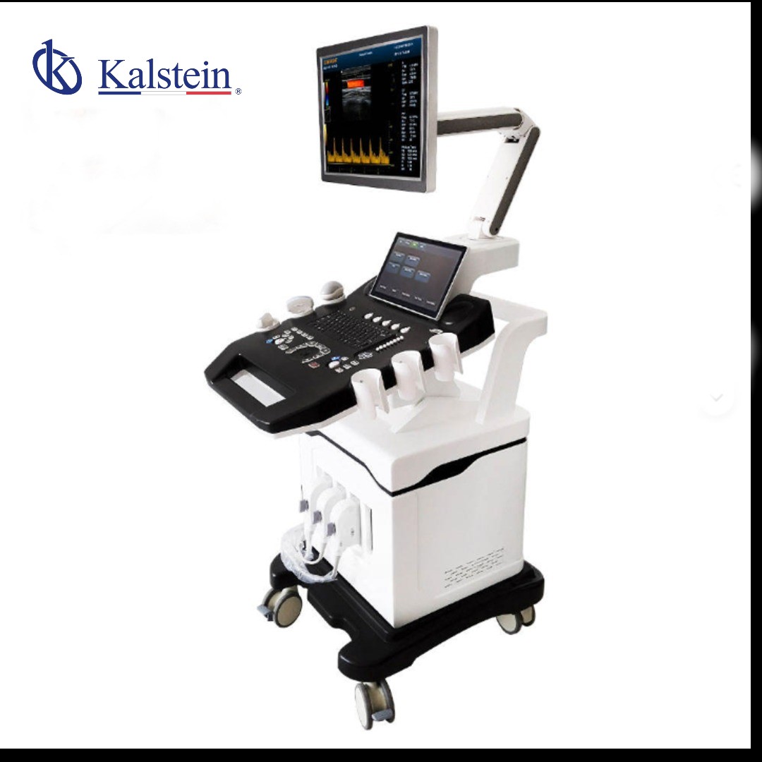
Color Doppler Ultrasound Cart with Wheels 3D Model YR05149
Color Doppler Ultrasound Cart with Wheels 3D Model YR05149 — equipo de laboratorio Kalstein con especificaciones técnicas, características avanzadas y soluciones profesionales certificadas para uso científico.
Market Price
The Color Doppler Ultrasound Cart, with its array of advanced features, is competitively priced in the market. While specific pricing might vary, potential buyers can expect a general market range between $7,500 to $8,300 USD for similar models. This price range reflects the comprehensive technology and reliability offered by this device. To get the best value, consider reaching out for a quote through Kalstein Plus.Frequently Asked Questions
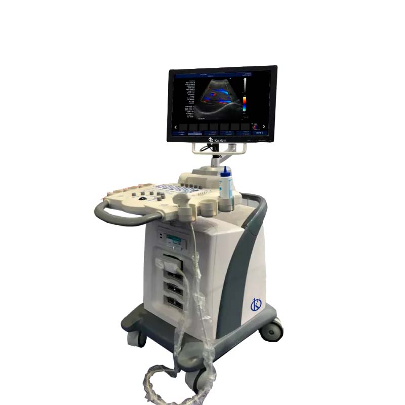
What kind of transducers does the YR05149 support? The YR05149 supports a range of transducers, including Convex, Linear, Intracavitary, and Microconvex probes, accommodating various clinical needs. Is the monitor adjustable? Yes, the 15" LED monitor has a ±90° rotation angle, allowing users to adjust the screen for optimal viewing in different environments. Can the ultrasound images be stored and exported? Absolutely. Images can be stored in various formats such as PNG, JPEG, BMP, and DICOM, and exported as AVI video files.Advantages and Disadvantages
One of the key advantages of the YR05149 model is its versatile imaging capabilities, thanks to features like multi-beamforming and dynamic tissue optimization technologies. These ensure high-quality images that improve diagnostic accuracy. The automatic measurements and compatibility with external devices enhance user efficiency. However, the array of features might be overwhelming for novice users, requiring a learning curve for maximizing all functionalities.Product Usage in the Field
The Color Doppler Ultrasound Cart is extensively used in hospitals and clinics for diverse applications, including obstetrics, cardiology, and internal medicine. Its mobility, facilitated by the cart with wheels, allows for easy transition between departments, making it an essential tool in fast-paced medical environments.Recommendations
To maintain optimal performance of the YR05149, it is vital to ensure regular software updates and calibrations. Also, familiarize yourself with the device's comprehensive user manual to utilize all features effectively. For any specific configuration assistance, engage with the online support function.Features
- 15" LED screen with multilingual support
- CF+B mode, PDI, DPDI, TDI, TSI capabilities
- Automatic measurements and quality assessment features
- Seamless DICOM 3.0 connectivity
- Integrated database and reporting functions
- Versatile connectivity with multiple ports
Technical Specifications
| Model | YR05149 | |||
| Connectivity/Media/Peripherals | ||||
| Transducer Ports | 4 | |||
| USB ports | 4 | |||
| HDD | 64GB (SSD), 120G/200GB SSD (optional) | |||
| Foot switch | USB | |||
| Ethernet port | 2(100Mb/1000Mb) | |||
| External screen | VGA, HDMI | |||
| Printer (Optional) | USB Printer, Digital Laser Printer, B/W Digital Thermal Printer | |||
| Printing area | Image, report, Image+report | |||
| Cine/Picture Memory | ||||
| Memory Cinema | 1200 frames (max) | |||
| Film Review Speed | 1, 2, 4, 8 | |||
| Cinema Review Loop | YES | |||
| Capture function | YES | |||
| DICOM connectivity | ||||
| DICOM3.0 Compliant | ||||
| 3D software | ||||
| Built-in 3D software | ||||
| Image Storage | ||||
| Storage Format | PNG, AVI, BMP, JPEG, DICOM | |||
| Export Video Format | AVI | |||
| Export Image Format | PNG, JPEG, BMP, DICOM | |||
| USB Flash Drive | ||||
| Technology | ||||
| Panoramic Imaging Technology Full Digital Signal Processing Technology Multi-Beamforming Technology Spot Reduction Technology Tissue Harmonic Imaging Technology Dynamic Tissue Optimization Technology Duplex and Triplex Synchronous Display Directional Power Doppler Imaging Parameters Imaging Parameters special tissue presets PW Auto Trace Update online CF+B mode on one screen Complex model imaging Automatic IMT measurements Virtual Convex Array Trapezoidal imaging | ||||
| Overall Performance | ||||
| Digital Broadband | 12288 channels | |||
| Beam Former | reprogrammable | |||
| Transmission voltage | Adjustable (15 steps) | |||
| Beamformer Frequency Range | 1~40MHz | |||
| Led Monitor | ||||
| Size (diagonal) | fifteen" | |||
| Contrast Ratio | 800: 01: 00 | |||
| Resolution | 1024×768 pixels | |||
| Brightness | 230 cd / m2 | |||
| Color Depth | 24 bit | |||
| Rotation Angle | ±90° | |||
| Gray levels | 256 | |||
| Imaging Performance | ||||
| Start Time (max.) | Average < 90 seconds | |||
| Preset Switching Time | Average < 1 second | |||
| Storage Time (Image to Disk) | Average < 0.5 seconds | |||
| Transducers | ||||
| Research | Convex array probe | Linear Array Probe | Intracavitary probe | Microconvex probe |
| Frequency | Center 3.5MHz | Central 7.5MHz | Center 6.5MHz | Center 4.0MHz |
| (2.0MHz to 10.0MHz) | (6.0MHz to 10.0MHz) | (5.0MHz to 9.0MHz) | (2.0MHz to 5.5MHz) | |
| Field of Play | 0.516mm | 0.352mm | 0.216mm | |
| Radio | 60mm | N/A | 10mm | |
Technical specifications
| Weight | 222 kg |
|---|---|
| Manufacturer | Kalstein |
Video Color Doppler Ultrasound Cart with Wheels 3D Model YR05149
Frequently asked questions
How to know the prices of Color Doppler Ultrasound Cart with Wheels 3D Model YR05149?
To know the price of Color Doppler Ultrasound Cart with Wheels 3D Model YR05149, please send us an email with your request through the contact form AQUI.
What are the delivery times for Color Doppler Ultrasound Cart with Wheels 3D Model YR05149?
Delivery time depends on stock availability and freight type (air or sea). In stock: air 15-30 days, sea 45-60 days. Out of stock: air 30-60 days, sea 60-90 days.
How to make a purchase of Color Doppler Ultrasound Cart with Wheels 3D Model YR05149?
You can buy by email ([email protected]), phone (+33 (0) 1 70 39 26 50) or through the official Kalstein website in your country.
How does the warranty work for Color Doppler Ultrasound Cart with Wheels 3D Model YR05149?
All Kalstein equipment comes with a 1-year warranty against manufacturing defects. The warranty does not cover damage from improper installation or misuse. See our «terms and conditions» AQUI.
Can I request a quote online for Color Doppler Ultrasound Cart with Wheels 3D Model YR05149?
Yes, you can request a quote for the Kalstein equipment you are interested in directly from our official website. Click AQUI.