Ultrasonic Color Doppler Diagnostic System: Practical Step-by-Step Guide 2026
The Ultrasonic Color Doppler Diagnostic System is a vital tool in modern medical imaging, utilizing advanced technology to provide real-time visualization of blood flow and organ structure. This guide aims to help laboratory professionals understand its applications, usage, techniques, and best practices in 2026.
What is it used for in 2026
In 2026, Ultrasonic Color Doppler Diagnostic Systems are primarily used for non-invasive imaging of blood flow and tissue structures within the body. They are essential in various medical fields including cardiology, obstetrics, gynecology, urology, and vascular studies. This technology helps in diagnosing conditions such as vascular disorders, cardiac abnormalities, and fetal health issues during pregnancy.
History and evolution of the technology
The development of ultrasonic imaging dates back to the early 20th century, but it wasn't until the 1960s that color Doppler technology emerged, allowing for dynamic visualization of blood flow. Over the decades, advancements in imaging resolution, software algorithms, and portable systems have transformed how Doppler ultrasound is applied in clinical practice, making it more accessible and effective for healthcare providers.
How to use it step by step
Using an Ultrasonic Color Doppler Diagnostic System involves several steps:
- Preparation: Ensure the machine is calibrated and properly connected. Clean the transducer and prepare the gel for better contact.
- Patient Positioning: Position the patient comfortably, ensuring the area of interest is accessible. Use pillows for support if necessary.
- Application of Gel: Apply a generous amount of ultrasound gel to the transducer to eliminate air pockets between the skin and the transducer.
- Transducer Placement: Place the transducer on the skin over the area of interest, angling it as needed to visualize blood flow.
- Settings Adjustment: Adjust the settings based on the area being examined (e.g., gain, depth, frequency) for optimal image quality.
- Image Acquisition: Capture images and video clips of the blood flow and structure, ensuring to record necessary measurements.
- Post-Procedure Care: Clean the gel from the patient's skin and review the captured data for completeness.
Best techniques and protocols
When utilizing Ultrasonic Color Doppler Systems, practitioners should adhere to the following best practices:
- Ensure consistent patient preparation to minimize variability in results.
- Use appropriate transducer frequency based on the depth and type of tissue being examined.
- Regularly calibrate and maintain the equipment to ensure accuracy.
- Utilize standardized measurement protocols to enhance reproducibility of results.
Practical applications by laboratory type
Ultrasonic Color Doppler Systems find applications in various laboratory settings:
- Cardiology Laboratories: Assessing heart conditions, measuring blood flow velocity, and diagnosing valvular heart diseases.
- Obstetrics and Gynecology: Monitoring fetal development, detecting anomalies, and evaluating blood circulation in the placenta.
- Vascular Laboratories: Evaluating peripheral arterial diseases, venous thrombosis, and other vascular conditions.
- Urology Laboratories: Assessing blood flow in renal arteries and examining testicular blood supply.
Regulations, standards and certifications
In the realm of medical imaging, adherence to regulations and standards is paramount. The devices must comply with the following:
- FDA regulations for medical devices in the United States.
- ISO 13485 standards for quality management systems in the manufacturing of medical devices.
- DICOM standards for medical imaging data and interoperability.
Comparison with alternative technologies
Compared to other imaging technologies such as MRI and CT scans, Ultrasonic Color Doppler offers several advantages:
- Non-invasive with no radiation exposure.
- Real-time imaging capabilities.
- Cost-effective and portable options available.
However, it may not provide the same level of detail for certain conditions that CT or MRI can reveal, especially in complex anatomical structures.
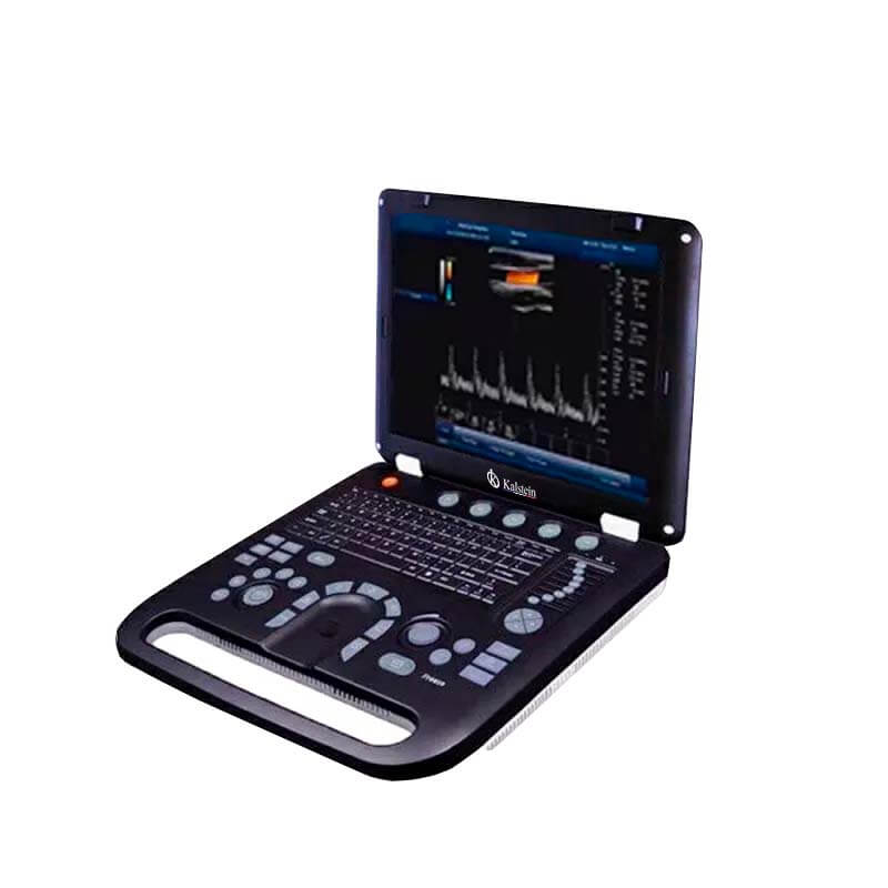
Comparison of available models
| Model | Best for | Key specs | Recommended use case |
|---|---|---|---|
| YR05148 | General diagnostic use | 15' LED display, multiple imaging modes | Standard examinations in various clinical settings |
| YR05149 | Mobile diagnostics | Portable with 4 transducer ports | On-the-go imaging for rural or emergency settings |
| YR05150 | Comprehensive vascular studies | 15'' color LED display, real-time Doppler function | In-depth vascular examinations |
| YR05151 | Advanced diagnostic capabilities | Multiple imaging modes, 4 transducer connectors | Complex cases requiring detailed imaging |
| YR05152 | Fetal health monitoring | Multilingual function, 4D capabilities | Obstetric examinations |
| YR05153 | Portable ultrasound scanning | 15' display, USB connectivity | Emergency and portable diagnostics |
Common mistakes and how to avoid them
Common pitfalls in the use of Ultrasonic Color Doppler systems include:
- Inadequate patient preparation, leading to suboptimal imaging results.
- Improper transducer handling, which can affect image quality.
- Neglecting equipment maintenance which can cause inaccuracies.
To avoid these, professionals should ensure thorough training, follow standard operating procedures, and regularly maintain equipment.
Maintenance, calibration and good practices 2026
Regular maintenance and calibration of Ultrasonic Color Doppler systems are crucial for optimal performance:
- Perform routine checks on software and hardware to ensure everything is functioning correctly.
- Calibrate the devices according to manufacturers’ guidelines to maintain accuracy.
- Keep the equipment clean, especially the transducer to ensure clear imaging.
Cost-benefit analysis 2026
In 2026, the cost of Ultrasonic Color Doppler systems varies based on features and specifications. However, their benefits often outweigh costs:
- High return on investment due to demand for non-invasive diagnostic procedures.
- Cost-effective compared to other imaging modalities like CT or MRI.
- Portable options reduce infrastructure costs for facilities.
Frequently asked questions
What should I consider when choosing an Ultrasonic Color Doppler System?
Consider factors such as imaging quality, portability, ease of use, and the specific types of examinations you will perform. Additionally, evaluate the system's compatibility with existing equipment.
How often should I calibrate my Ultrasonic Color Doppler equipment?
Calibrate your equipment at least once a year, or more frequently if there are significant changes in performance or after any repairs.
What are the main limitations of Ultrasonic Color Doppler technology?
Limitations include its reduced effectiveness in imaging deeper structures and potential operator dependency affecting outcomes.
Can I use the system for both obstetrics and vascular studies?
Yes, Ultrasonic Color Doppler Systems are versatile and can be utilized for various applications including obstetrics and vascular studies, depending on the model's specifications.
What is the expected lifespan of an Ultrasonic Color Doppler machine?
With proper maintenance and care, these machines can last over 10 years, but technology updates may necessitate earlier replacements.
Is training required to operate these systems effectively?
Yes, proper training is crucial for effective operation and ensuring accurate diagnostics using Ultrasonic Color Doppler systems.
Where can I find support for my Ultrasonic Color Doppler system?
Most manufacturers provide customer support and technical assistance for their devices. Additionally, many online resources and user communities can offer guidance.
If you are looking for a fusion of innovation and quality, you have come to the right place. At Kalstein, we offer you the luxury of exploring our exclusive catalog of laboratory equipment. We manufacture every device to the highest standards of excellence. Our intuitive and seamless online purchasing channels are designed for your convenience, securing the most competitive prices. Hesitate no longer — we bring science to life, it is time to become part of our community.